
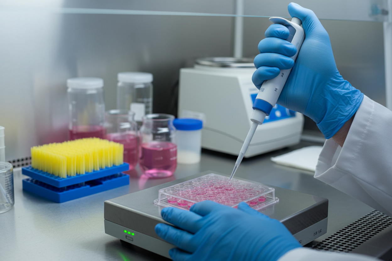
Gene delivery is one of those lab steps that looks simple on paper but behaves differently every time you run it. You follow the same protocol, use the same plasmid, the same cells, and still end up with wildly different expression levels, low titers, or a plate full of stressed cells.
This is where PEI transfection stands out. It’s one of the most widely used non-viral gene delivery methods because it’s scalable, cost-effective, and adaptable across cell types. But it’s also highly sensitive to small changes in experimental conditions. A slightly different cell density, a longer incubation time during complexation, or a small shift in ratio can turn a great transfection into a complete failure.
In this post, we’ll break down what actually influences PEI performance, what most protocols overlook, and how to systematically optimize without relying on trial-and-error guesswork.
Why PEI Transfection Works (and Why It Sometimes Doesn’t)
PEI is a polycation (positively charged polymer) that binds DNA (negatively charged) to form compact DNA–polymer complexes called polyplexes. These polyplexes help DNA enter cells via endocytosis. Once inside, PEI’s buffering capacity supports endosomal escape—often described as the “proton sponge” effect—allowing the DNA to be released and expressed.
The problem is that PEI is effective partly because it’s aggressive. If the complexes are too positively charged or too concentrated, cells experience stress, reduced viability, and inconsistent expression.
So PEI success is always a balance between:
- Efficient uptake
- Endosomal escape
- Minimal toxicity
The Real Control Knob: DNA:PEI Ratio (N/P Ratio)
If there’s one variable that controls the entire PEI outcome, it’s the N/P ratio—the ratio of PEI nitrogen groups to DNA phosphate groups. It determines the overall charge of the polyplex and influences:
- complex stability
- aggregation behavior
- uptake efficiency
- toxicity
A common mistake is assuming “more PEI = better transfection.” In reality, adding PEI past the optimal point often increases uptake slightly but causes enough toxicity to reduce net expression.
A better strategy is to run a matrix like:
- N/P 5, 10, 15, 20
- across multiple DNA amounts (e.g., 0.5, 1, 2 µg per million cells)
Then measure both:
- expression (GFP/luciferase/ELISA)
- viability (24h, 48h, 72h)
This reveals the best “high expression + high viability” zone instead of chasing expression alone.
DNA Quality: The Silent Efficiency Killer
When PEI transfection fails, many people immediately blame the reagent or ratio. But one of the most common hidden issues is DNA quality, especially endotoxin contamination.
Even low endotoxin levels can:
- reduce transgene expression
- activate stress pathways
- increase PEI-associated cytotoxicity
- cause inconsistent results between runs
If your DNA is:
- cloudy
- stringy
- low A260/280
- from a rushed prep
…it can sabotage even a perfectly optimized PEI protocol.
Cell Health Isn’t Optional
PEI does not “fix” unhealthy cells—it punishes them.
For reproducible results, cells should be:
- actively dividing (log phase)
- high viability (ideally ≥95%)
- not over-passaged
- not stressed from overconfluency, starvation, or contamination
One of the biggest reproducibility issues in PEI workflows is simply different cell conditions between experiments. If cells are even slightly stressed, they become more sensitive to PEI and show lower expression even at the same ratio.
Complexation Timing: Small Changes, Big Consequences
Complex formation isn’t instantaneous. PEI and DNA need time to form stable polyplexes.
A typical optimal range is:
- 10–30 minutes at room temperature
If complexation time is:
- too short → incomplete complex formation, poor uptake
- too long → aggregation, increased toxicity, lower expression
Also, once you identify a working complexation time, keep it fixed. Timing inconsistencies create variability that looks like “random transfection behavior” but is actually protocol drift.
Avoid the “Invisible Interference” in Media
Another overlooked issue is what’s in the buffer/media during complex formation.
Things that interfere with PEI–DNA complexation:
- serum proteins
- antibiotics/antimycotics
- phosphate buffers
These components can destabilize complexes or increase toxicity after uptake. A simple improvement is to form complexes in a compatible serum-free medium, then restore complete medium later depending on the sensitivity of your cell line.
Post-Transfection Care: The Step People Skip
After transfection, leftover free PEI in the medium can continue to stress cells. Some cell lines tolerate it. Others collapse.
A practical rule:
- if toxicity is high → replace media 4–6 hours post-transfection
- if cells are robust → test both with and without replacement
The goal isn’t to follow a universal “must change media” rule—it’s to build a workflow that fits your cell line and output (protein expression, AAV, LV, etc.).
The Most Reliable Way to Optimize PEI Transfection
If you want consistent results, don’t optimize randomly. Optimize like an engineer:
- Start with a reporter (GFP or luciferase)
- Test DNA amount range
- Test N/P ratio range
- Fix complexation buffer and time
- Standardize seeding density and viability
- Run a time course to identify peak expression window
This approach turns PEI transfection from “lab luck” into a controlled, scalable system.
If you want a structured protocol-style guide that expands these optimization variables into a step-by-step framework, you can reference this resource: pei transfection.



More Stories
Voyage of the Cosmic Echoes: A Sci-Fi Odyssey Beyond the Event Horizon
The Next Dimension: Imagining the Cities of Tomorrow
The Quantum Echoes of Tomorrow